

Ultrasound (USG) Whole Abdomen
An ultrasound examination of the whole abdomen, commonly referred to as “USG Whole Abdomen,” is a non-invasive and widely used medical imaging procedure. It involves the use of high-frequency sound waves to create detailed images of the structures within the abdomen. This diagnostic technique is valuable for assessing various abdominal organs, tissues, and potential abnormalities without the need for radiation exposure.
Purpose of the Procedure:
USG Whole Abdomen is performed for a variety of diagnostic purposes, including:
- Organ Evaluation: The procedure allows medical professionals to assess the condition, size, shape, and position of vital organs such as the liver, gallbladder, spleen, pancreas, kidneys, and bladder.
- Detection of Abnormalities: USG can help identify abnormalities such as tumors, cysts, stones, and masses within the abdominal cavity.
- Evaluation of Blood Flow: It can provide information about blood flow within organs, helping diagnose conditions like liver cirrhosis or kidney vascular issues.
- Assessment of Infections or Inflammation: Infections or inflammation in organs like the liver, gallbladder, or appendix can be visualized through ultrasound imaging.
- Monitoring Pregnancy: For pregnant individuals, a USG Whole Abdomen can track fetal growth, amniotic fluid levels, and the overall health of the developing fetus.
Procedure Overview:
During the procedure:
- Preparation: Typically, no extensive preparation is required. Patients might be asked to fast for a few hours before the procedure, especially if the examination involves the gallbladder.
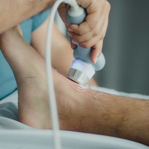
- Positioning: The patient lies on an examination table. A water-based gel is applied to the abdomen to facilitate better sound wave transmission.
- Transducer Application: A transducer, a small handheld device that emits and receives sound waves, is gently moved across the abdomen by a trained sonographer or radiologist.
- Image Formation: As the transducer sends sound waves into the body, they bounce off organs and tissues, creating echoes. The echoes are captured by the transducer and processed by a computer to generate real-time images on a monitor.
- Assessment: The healthcare professional will interpret the images in real time, looking for any anomalies or abnormalities. Measurements and observations are noted.
- Completion: Once the necessary images are captured, the gel is wiped off, and the procedure is complete.
Benefits and Limitations:
Benefits of USG Whole Abdomen include its non-invasive nature, real-time imaging capabilities, absence of radiation, and suitability for various patient groups. However, its effectiveness may be limited in cases where air or gas obstructs the sound waves, limiting the view of certain organs or areas.


Reviews
There are no reviews yet.